News
Neurosoft Bioelectronics: brain-machine interfaces that are softer, less invasive, and richer in data

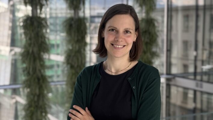
© Roger do Minh
Brain-machine interfaces could transform how certain neurological conditions are diagnosed and treated. But for devices to be usable over the long term, one question remains central: how to record brain activity with enough precision while limiting the impact on brain tissue? That is the question Neurosoft Bioelectronics is working on – an EPFL spin-off based at Campus Biotech in Geneva.
A non-penetrating architecture
Neurosoft Bioelectronics is developing a brain-computer interface built on soft electrodes designed to rest on brain tissue rather than pierce it. In practice, the company relies on soft, stretchable electrodes – up to 1,000 times more flexible than existing neural interfaces – arranged on implants just a few square centimetres in size. Placed on the surface of the cortex rather than inserted into it, they provide full cortical access without penetrating the tissue, unlike the needle-like electrode arrays used in some current brain-computer interfaces, which offer greater local precision at the cost of higher tissue risk over time.
This approach has another payoff: by making cortical recording safer and broader in scope, it yields richer data – the kind AI models need to learn how to translate the brain’s electrical activity into movement or speech.
A safety standard that goes beyond regulation
For Nicolas Vachicouras, CEO and co-founder, this is not simply a matter of performance: it is also a safety standard, particularly for devices meant to be worn continuously, sometimes for decades. Beyond mandatory regulatory testing, he applies a personal criterion before signing off on any device:
« There’s always the regulatory test – that’s mandatory. But there’s another test, too, one I call the “mother test”: would I be comfortable putting this device in my own mother’s head? Regulation exists to provide certainty, but it can also, in some cases, hold back technologies that are actually safe. It’s a difficult balance, but one worth getting right ».
That standard plays out concretely in the indications Neurosoft is pursuing: beyond epilepsy, its most advanced indication to date, the company is also exploring motor impairments and severe tinnitus – applications in which the implant is meant to remain in place for years.
An award that confirms a broader momentum
This approach was recently recognized at the 2026 Grands Prix de l’économie, where Neurosoft Bioelectronics received the Innovation Award, presented on September 17, 2026 before an audience of some 600 entrepreneurs and business leaders at HEAD – Geneva University of Art and Design. What the jury recognized was the ability of soft, minimally invasive brain interfaces to decode brain activity at scale while leaving brain tissue intact.
This recognition fits into a trajectory already marked by concrete results: in May 2026, Neurosoft closed a USD 7.5 million funding round, bringing its total funding to more than USD 20 million, with investor confidence anchored in part by a portfolio of more than 25 patents. The company, incubated at Campus Biotech with support from the Wyss Center before expanding to New York, continues to manufacture its electrodes there.
Toward US regulatory clearance
What comes next is already taking shape on the clinical and regulatory front: Neurosoft plans to expand its clinical trials across Europe and the United States, while seeking US regulatory clearance for its first product, targeting epilepsy – its most advanced indication.