 News
News
Restless nature of human spinal cord, non-invasive imaging reveals
 Partner
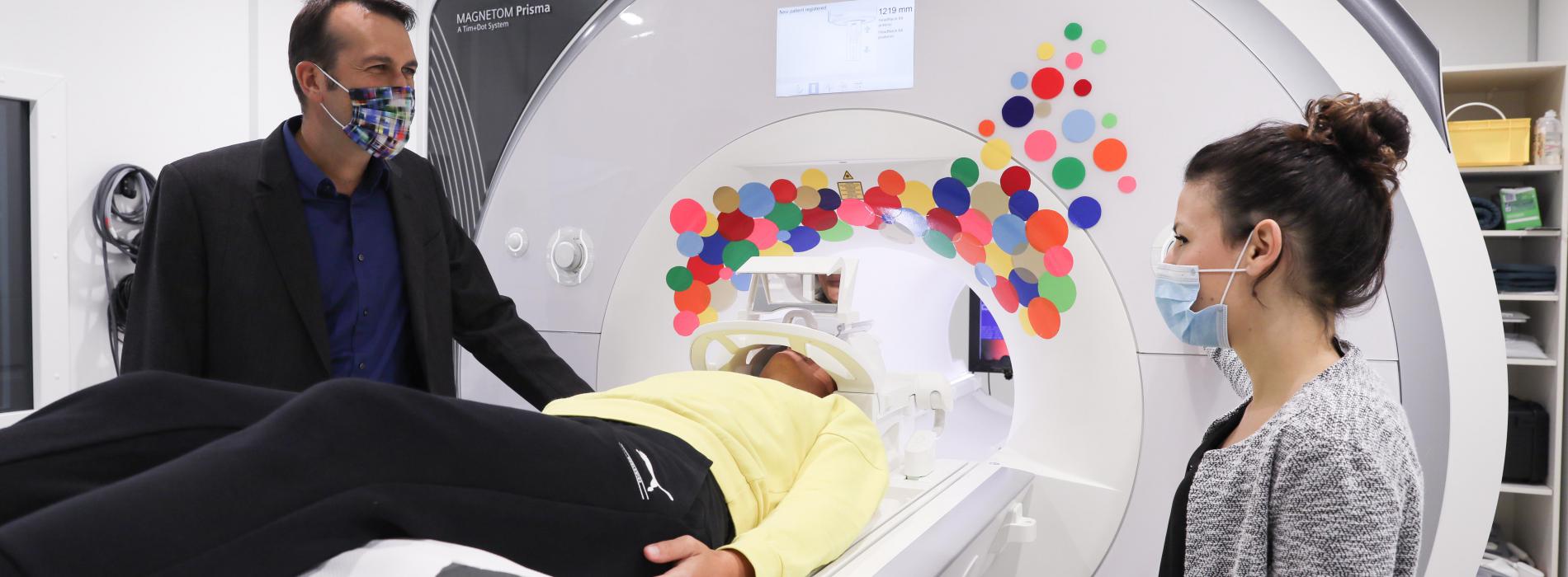
© 2020 EPFL / Dimitri Van De Ville (left) and Nawal Kinany (right) with a healthy subject, at Campus Biotech.
EPFL scientists have developed a non-invasive technique for unraveling the complex dynamics generated by spinal cord circuits to unprecedented detail, a first in functional magnetic resonance imaging that may one day help diagnose spinal cord dysfunction or injury.
The spinal cord roughly looks like a long tube, with a diameter of only 1.5 cm, and yet this crucial part of the nervous system is essential for controlling how our arms and legs move, for giving us our sense of touch as well as a notion of where our bodies are in space.
How does this seemingly simple structure support complex behaviors? To answer this question, scientists have longed for a way to observe the spinal cord’s function in vivo. Until recently, they needed to resort to animal studies, but the advent of fMRI (functional magnetic resonance imaging) is now providing a new window into the richness of spinal cord signals, directly in humans.
Now, scientists at EPFL have combined tailored protocols for spinal cord fMRI with advanced analysis techniques, in order to disentangle these signals and clearly view the spinal cord in action. Tested on 19 healthy subjects, the scientists obtained unprecedented views of the spinal cord’s functional architecture and showed for the first time just how dynamic the spinal cord is, even for subjects at rest. The results are published in today’s issue of Neuron.
“One of the main challenges about observing spinal cord function is getting rid of noise from the rest of the subject’s body, like breathing, the heartbeat, or simply seeing beyond the surrounding vertebral bones,” explains Nawal Kinany, first author of the study. “We managed to decompose spontaneous spinal activity into meaningful networks, with a level of neuroanatomical detail that had never been reached before.”
The study was done at Campus Biotech in collaboration with Silvestro Micera who is the Bertarelli Foundation Chair in Translational Neuroengineering at EPFL and Professor of Bioelectronics at Scuola Sant’Anna in Pisa, Italy, as well as with Dimitri Van De Ville who leads EPFL’s Medical Imaging Processing Lab and is also affiliated with the Department of Radiology and Medical Informatics of the Geneva University.
From the perspective of the subject, one simply has to lie down in an fMRI scanner and remain immobile throughout the scan, typically around 10 minutes. The images resulting from the scan are then analyzed to provide a 4-dimensional view – through space and time – to view the dynamics of spinal circuits within the anatomy of the subject.
“These results are clear evidence that spinal resting-state activity is richly organized and thus must bear physiological relevance beyond what was assumed so far,” explains Van De Ville.
Given its central position at the interface between the brain and the rest of body, the spinal cord is a key player in all human behaviour. The scientists targeted the cervical level because of its involvement in controlling arm and hand muscles. Their approach could help understand how spinal circuits are orchestrated to support the wide range of movements we perform in our everyday life.
“Only a deeper understanding of human motor control can allow for the development of more effective neurorehabilitation approaches. Our new method provides a very important tool in this direction,” furthers Micera.
Tested for now on healthy subjects, the scientists believe that these new protocols will one day be a valuable tool for evaluating the status of dysfunctional or injured spinal cord circuitry, which could promote the development of targeted therapies that rebalance spinal activity or optimally harness the spared connections.
https://actu.epfl.ch/news/restless-nature-of-human-spinal-cord-non-invasiv-3/