The aim of the Imaging and Microscopy facility is to offer both technical support and innovation in imaging technology to the scientific community in academia and in industry.
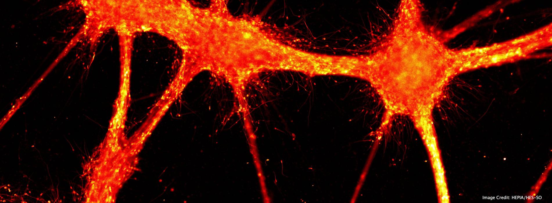
The facility includes state of the art light microscopy instrumentation, ranging from slide scanner, and confocal laser scanning and light-sheet microscopy to advanced software for image analysis.
The availability of cutting-edge tools such as X-clarity, custom light-sheet systems for whole brain acquisition and 4D post-processing software, together offer to neuroscientist unique opportunities to explore uncovered boundaries.
Equipment:
Widefield Microscopy
Leica DM4000B
Olympus VS-120 Slide scanner
Confocal Microscopy
Zeiss LSM880 + Airy fast module
Leica TCS SPE-II DM5500 RGB
Lightsheet Microscopy
Clarity Optimized Ligthsheet Microscope (COLM) (custom microscope)
MesoSPIM (custom microscope)
Image analysis workstation equipped with Arivis and Imaris software
This platform is under the scientific supervision of Professor Grégoire Courtine (EPFL/CNP-BMI), Professor Diego Ghezzi (EPFL/CNP).
The Imaging and Microscopy facility, at Campus Biotech embeds the new Advanced Lightsheet Imaging Center (ALICe).
The imaging and microscopy team is composed by Laura Batti, Stéphane Pagès and Ivana Gantar. For more information please, contact us: microscopy@wysscenter.ch
 Research Team
